OBJECTIVE: To determine the relationships between conventional and segmentation-derived optical coherence tomography (OCT) retinal layer thickness measures with intracranial volume (a surrogate of head size) and brain substructure volumes in multiple sclerosis (MS).
PARTICIPANTS: A total of 84 patients with MS and 24 healthy control subjects.
MAIN OUTCOME MEASURES: High-definition spectral-domain OCT conventional and automated segmentation-derived discrete retinal layer thicknesses and 3-T magnetic resonance imaging brain substructure volumes.
RESULTS: Peripapillary retinal nerve fibre layer as well as composite ganglion cell layer + inner plexiform layer thicknesses in the eyes of patients with MS without a history of optic neuritis were associated with cortical gray matter (P = .01 and P = .04, respectively) and caudate (P = .04 and P = .03, respectively) volumes. Inner nuclear layer thickness, also in eyes without a history of optic neuritis, was associated with fluid-attenuated inversion recovery lesion volume (P = .007) and inversely associated with normal-appearing white matter volume (P = .005) in relapsing-remitting MS. As intracranial volume was found to be related with several of the OCT measures in patients with MS and healthy control subjects and is already known to be associated with brain substructure volumes, all OCT-brain substructure relationships were adjusted for intra-cranial volume.
CONCLUSIONS: Retinal measures reflect global central nervous system pathology in multiple sclerosis, with thicknesses of discrete retinal layers each appearing to be associated with distinct central nervous system processes. Moreover, OCT measures appear to correlate with intracranial volume in patients with MS and healthy control subjects, an important unexpected factor unaccounted for in prior studies examining the relationships between peripapillary retinal nerve fibre layer thickness and brain substructure volumes.
The study looks at the thickness of the retina, which is known to thin in multiple sclerosis. This study correlates the thickness of the retina and the volume of the brain and indicates that there is some correlation so the more the retina thins the lower the volume of the brain is. It is quick and easier to measure the thickness of the eye and it is not surprising that the more disease activity that cause damage to the brain could also result in damage to the optic nerve that results in damage of the eye There is some correlation of the imaging some areas of the eye with certain brain areas. Can you use the imaging of the eye to say what is going on in the brain? Well the partial problem is the relationships are not absolute. However Team G are using this as an outcome in an clinical trial to assess the effect of a neuroprotective drug.

If you have a demyelinating attack in the optic nerve it damages the axons and this can die back and kill the nerve cell whose nucleus is in the retina
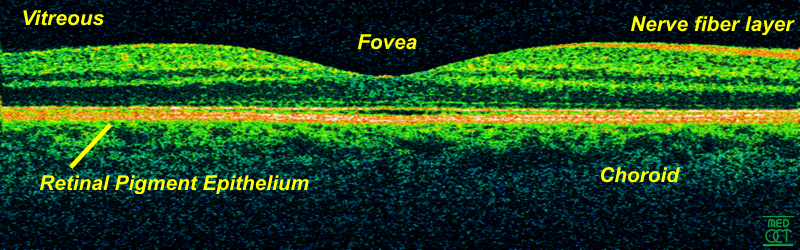
 Just so you can see here is MS in a human Eye, well it is actually in the optic nerve .